UNIVERSIDAD AUTONOMA DE BARCELONA DEPARTAMENTO DE PEDIATRIA, OBSTETRICIA Y GINECOLOGÍA Y MEDICINA PREVENTIVA UTILIDAD DEL MAPA LINFATICO EN EL
|
|
|
- Joaquín Mora Marín
- hace 8 años
- Vistas:
Transcripción
1
2 UNIVERSIDAD AUTONOMA DE BARCELONA FACULTAD DE MEDICINA DEPARTAMENTO DE PEDIATRIA, OBSTETRICIA Y GINECOLOGÍA Y MEDICINA PREVENTIVA UTILIDAD DEL MAPA LINFATICO EN EL TRATAMIENTO QUIRURGICO DE LOS ESTADIOS PRECOCES DEL CANCER DE CERVIX Tesis doctoral presentada por el licenciado Josep Mª Martínez i Palones para optar al grado de Doctor en Medicina y Cirugía.

3 A mi esposa. A mis hijos Gemma, Alex y Víctor, por la paciencia que ha tenido conmigo.

4
5 10

6 11

7 12
8 13
9 14
10 Gynecologic Oncology 96 (2005) Total laparoscopic radical hysterectomy with intraoperative sentinel node identification in patients with early invasive cervical cancer Antonio Gil-Moreno a, *, Berta Díaz-Feijoo a, Isabel Roca b, Oriol Puig a, María A. Pérez-Benavente a, Ignacio Aguilar a, José M. Martínez-Palones a, Jordi Xercavins a a Unit of Gynecologic Oncology, Department of Obstetrics and Gynecology, Hospital Materno-infantil Vall d Hebron, Spain b Service of Nuclear Medicine, Hospital General Vall d Hebron, Autonomous University of Barcelona, Barcelona, Spain Received 20 June 2004 Abstract Objectives. To describe the feasibility and results of total laparoscopic radical hysterectomy with intraoperative sentinel lymph node identification in patients with early cervical cancer. Methods. Between March 2001 and October 2003, 12 patients with FIGO stage IA 2 (n = 1) or IB 1 (n = 11) cancer of the cervix underwent surgical treatment through the laparoscopic route. All patients underwent a laparoscopic sentinel node identification with preoperative lymphoscintigraphy (technetium-99 m colloid albumin injection around the tumor) and intraoperative lymphatic mapping with isosulfan blue dye and a laparoscopic gamma probe followed by systematic bilateral pelvic lymphadenectomy and laparoscopic type II (n = 5) or type III (n = 7) hysterectomy. Results. A mean of 2.5 sentinel nodes per patient (range 1 4) was detected, with a mean of 2.33 nodes per patient by gamma probe and a mean of 2 per patient after blue injection (combined detection rate 100%). The most frequent localization of the nodes was the interiliac region. Histopathologic examination of sentinel nodes including cytokeratin immunohistochemical analysis did not show metastasis. Microscopic nodal metastases were not found. The mean number of resected pelvic nodes was 18.6 per patient (range 10 28). The operation was performed entirely by laparoscopy in all patients and no case of laparotomy conversion was recorded. The mean duration of operation was 271 min (range ), with a mean blood loss of 445 ml (range ), and a mean length of stay of 5.25 days (range 3 10). No major intraoperative complications occurred. After a median follow-up of 20 months (range 5 34), all patients are free of disease. Conclusions. This study shows the feasibility of the combination of laparoscopic intraoperative sentinel node mapping and laparoscopic radical surgery in the context of minimally invasive surgery for the management of patients with early cervical cancer. D 2004 Elsevier Inc. All rights reserved. Keywords: Laparoscopic radical hysterectomy; Cervical cancer; Laparoscopic surgery; Pelvic lymphadenectomy; Sentinel lymph node; Surgical morbidity Introduction Lymph node status is a major prognostic factor for patients with cervical carcinoma and is a decision criterion for adjuvant therapy. The concept of sentinel node mapping * Corresponding author. Unit of Gynecology Oncology, Department of Obstetrics and Gynecology, Hospital Materno-infantil Vall d Hebron, Passeig Vall d Hebron , E Barcelona, Spain. Fax: addresses: agil@cs.vhebron.es, antonioimma@yahoo.es (A. Gil-Moreno). was proposed by Canabas for the management of patients with penile cancer using lymphangiograms performed via the dorsal lymphatics of the penis [1,2]. The sentinel lymph node is defined as the first draining lymph node of an anatomical region, so that a histologically negative sentinel lymph node would predict the absence of tumor metastases in the other non-sentinel lymph nodes. Detection of sentinel lymph node status has become accepted for melanoma and breast cancer [3 5] and is a promising staging technique for patients with vulvar cancer [6,7]. In patients with early cervical cancer, pelvic or paraaortic lymph node metastases are detected in only 8% of women /$ - see front matter D 2004 Elsevier Inc. All rights reserved. doi: /j.ygyno

11 188 A. Gil-Moreno et al. / Gynecologic Oncology 96 (2005) with stage IA 2 and in 26% of women with stage IIA no bulky [8]. Thus, many of these patients derive no benefit from pelvic lymphadenectomy. On the other hand, this procedure may increase morbidity, especially when postoperative radiotherapy is required. Intraoperative detection of sentinel lymph node status may have significant implications from a clinical management point of view. If the node is positive for tumor metastasis, a radical hysterectomy could be omitted, and adjuvant chemoradiation is commonly given [9 11]. Different studies have demonstrated the relevance of sentinel node identification using blue dye alone [12], radiocolloid alone [13], or a combination of the two reagents [9,10,14], with laparotomy [13,14] or using a laparoscopic procedure [9,10,11,12,14]; laparoscopy is associated with the potential benefits of decreased discomfort and shorter hospital stays. Radical hysterectomy (class II or class III) [15] by the vaginal or the abdominal route is the surgical procedure used in treating women with cervical cancer. However, a vaginal [16] or a laparoscopic approach [10,14,17] in radical surgery for cervical carcinoma has been shown to be feasible in accordance with the standards of gynecologic oncology. The purpose of this study was to describe our initial experience with total laparoscopic radical hysterectomy for early cervical cancer (FIGO stages IA 2, IB 1, IIA no bulky) combined with laparoscopic intraoperative sentinel lymph node identification. Patients and methods Between March 2001 and October 2003, 12 nonconsecutive patients with FIGO stage IA 2 (n = 1) or IB 1 (n = 11) cancer of the cervix were candidates for total laparoscopic radical hysterectomy and intraoperative sentinel node identification. The diagnosis of cervical carcinoma was made either on cervical punch biopsy or after a cone biopsy. Intraoperative identification of sentinel lymph node was performed entirely by the laparoscopic route with preoperative lymphoscintigraphy (technetium-99 m-labeled nanocolloid) and intraoperative lymphatic mapping with isosulfan blue dye followed by systemic bilateral pelvic lymphadenectomy and radical hysterectomy by the laparoscopic approach. Patients were informed of risks and complications and provided written informed consent for possible need to convert to open surgery. Exclusion criteria for the sentinel node identification procedure were bulky disease which made injection in normal cervical tissue impossible and prior chemotherapy or pelvic radiotherapy. Inclusion criteria for total laparoscopic radical hysterectomy were a tumor size b4 cm and no evidence of node involvement in imaging studies (MRI scans). A body mass index (BMI) V35 kg/m 2 was also required. Patients at stage IA 2 and patients at stage IB 1 with size of the primary lesion V2 cm underwent a type II procedure, and patients at stage IB 1 with tumors measuring N2 cm a type III procedure as defined by Piver et al. [15]. Combined general and epidural anesthesia was used in every case. Patients were administered perioperative antibiotics, cefazoline 2 g intravenously, and low molecular weight enoxaparin (40 mg/24 h subcutaneously). Sentinel node identification procedure The day before surgery, the cervix viewed with a speculum was superficially injected in each quadrant around the tumor at 3, 9, 6, and 12 o clock positions with depots of technetium- 99 m-labeled nanocolloid (Albu-resR, Pharmaceutical Nycomed Amersham, Bruanschweig, Germany), each containing approximately 10 mbq of tracer in 0.2 ml saline, using a 27-gauge hypodermic needle. Immediately after injection and removal of the speculum, dynamic lymphoscintigraphy was performed using a gamma camera. Additional static images were taken after the dynamic study and about 2 3 h after injection to identify the sentinel node. At the time of surgery, after induction of general anesthesia and speculum viewing of the cervix, approximately 2 4 ml of isosulfan blue dye (Lymphazurin 1%, US Surgical Co., Norwalk, CT) was injected superficially around the tumor at the same locations as the tracer (depth of injection cm). For the laparoscopic procedure, a four-trocar transperitoneal approach was used. The laparoscope was introduced via an 11-mm umbilical trocar with a direct-puncture technique, and three other trocars were inserted with the patient in a 258 to 308 Trendelenburg position. Two accessory 12- and 5-mm trocars were then placed under direct visualization in the iliac fossae, and one 5-mm accessory trocar at the midline between the umbilicus and the left subcostal margin. The abdominal cavity was inspected and the retroperitoneal spaces were opened. The sentinel node was detected using a laparoscopic gamma probe NavigatorR (AutoSuture, Norwalk, CT) or NeoprobeR (Ethicon Endo-Surgery, Cincinnati, OH). Nodal tissue was examined visually for blue staining (Fig. 1) and audible signs above background. If count were at least 10- fold above background levels, the node was considered sentinel. Sentinel nodes were labeled as blue, hot (radioactive), or blue/hot. All sentinel nodes were recorded by their relative position to the major pelvic vessels. Each blue and/or hot lymph node was removed separately in endoscopic bags (Endo-Catch Gold 10 mm, AutoSuture, Norwalk, CT) and submitted for pathologic examination. Systematic, laparoscopic, bilateral pelvic lymphadenectomy from the common iliac artery to the inferior boundary of the circumflex iliac vein was performed after the sentinel node procedure (Fig. 2). Laparoscopic radical hysterectomy With the same disposition of the four trocars, the paravesical, pararectal, rectovaginal, and vesicovaginal spaces were developed, thereby delineating the uterosacral 16

12 A. Gil-Moreno et al. / Gynecologic Oncology 96 (2005) encountered bilaterally, the vagina was transected with monopolar coagulation, and the specimen was removed vaginally (Fig. 3). The vaginal cuff was then closed laparoscopically with 2 0 polyglactin (Vicryl, Ethicon GmbH) continuous suture, using the KOH colpotomizer (CooperSurgical, Trumbull, CT) for obturation of the vagina to avoid pneumoperitoneum air leakage (Fig. 4). After completing the vaginal procedure, we checked for hemostasis and intact ureters, and the peritoneal cavity was thoroughly irrigated and lavaged. A suprapubic catheter was placed. The catheter was maintained until residual urine volumes b100 ml were regularly obtained for a minimum of 2 weeks (range 2 7 weeks). Pathological evaluation Fig. 1. Identification of a blue-positive sentinel lymph node. ligaments and the parametria. Uterine vessels were identified and cut at their origin from the hypogastric vessels with vascular clips in the first cases and using the harmonic scalpel (Ultracision Harmonic Scalpel, Ethicon Endo-Surgery) more recently. The parametrial tissue and the uterosacral ligaments were transected with the Endo-GIA stapler (ATW35, TSB45 Endo-Path, Ethicon Endo-Surgery) in the first cases and using the harmonic scalpel more recently. In patients in which ovarian function was maintained, the ovaries were transposed laterally to the paracolic gutters and fixed securely to the abdominal wall. Dissection of the ureteral tunnel, urinary bladder, and ureteral separation was accomplished with monopolar coagulation using a blunt dissector and with the aid of the Ultracision. Resection of the proximal paracervical tissues was performed with the flexible Endo-GIA stapler or the harmonic scalpel. At the point, the vaginal fornices were Sentinel lymph nodes were visually inspected by the pathologist. If gross metastatic disease was visible, simple sections were submitted. Normal-appearing sentinel nodes were cut perpendicularly to the long axis (one slide per each 0.2-mm section) and submitted for routine hematoxylin and eosin (H&E) staining. Negative H&E sections were prepared for wide spectrum cytokeratin immunohistochemical analysis (CK-88). Data of each individual patient were registered prospectively using an Access and Excel database (Microsoft Inc.). Results Patients had a mean age of 43 years (range 35 57), with a mean weight of 61 kg (range kg), and mean BMI of 24.9 kg/m 2 (range kg/m 2 ). History of previous surgery was recorded in two patients (cesarean section in 1, laparoscopic tubal ligation in 1). None of the patients had Fig. 2. Left pelvic lymphadenectomy. External iliac artery, 1; external iliac vein, 2; internal iliac artery, 3; obturator nerve, 4; genitofemoral nerve, 5; uterine artery, 6. Fig. 3. Surgical specimen after total laparoscopic radical hysterectomy. 17
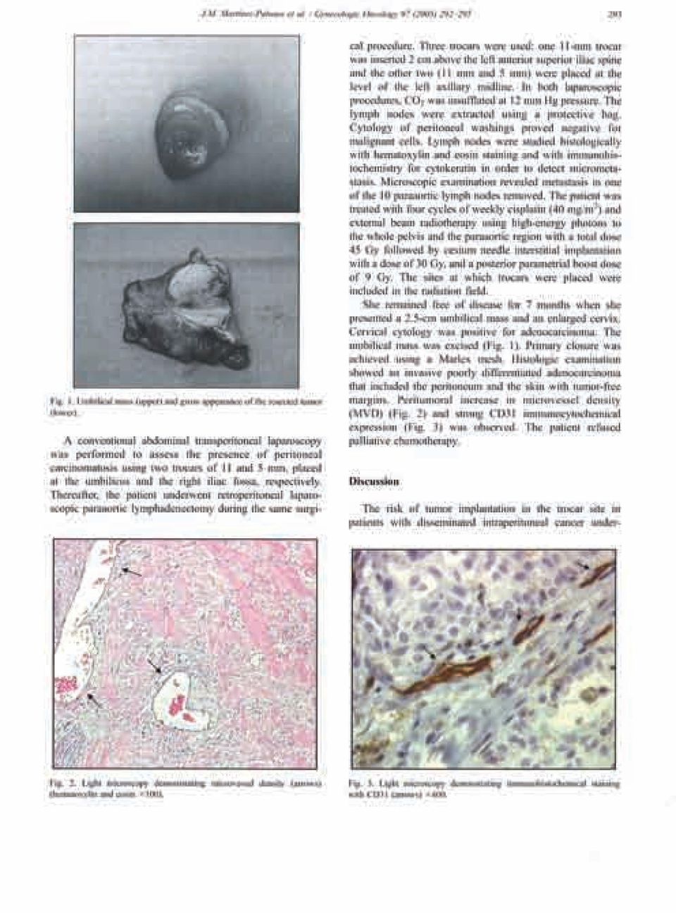
. Pathological evaluation Fig. 1.")
13 190 A. Gil-Moreno et al. / Gynecologic Oncology 96 (2005) Fig. 4. Suture of the vaginal cuff through the laparoscopic route. MRI evidence of lymph node involvement. Histological types included squamous cell carcinoma in 67% of patients and adenocarcinoma in 33%. Vascular invasion was found in five patients. The surgical margins were free of disease in all cases. The mean tumor size in patients with FIGO stage IB 1 was 2.36 cm. Clinical, intraoperative, and pathologic characteristics of the study population are presented in Table 1. The operation was performed entirely by laparoscopy in all patients (type II hysterectomy, n = 5; type III hysterectomy, n = 7) and no patient required conversion to laparotomy. Ovarian transposition was performed in eight premenopausal patients. The mean duration of operation was 271 min (range min). The mean intraoperative blood loss was 445 ml (range ml). The mean length of hospital stay was 5.25 days (range 3 10 days). There were no intraoperative complications. Early postoperative complications occurred in two patients and included urinary tract infection in one and blood transfusion in a patient with postoperative symptoms of anemia and a serum hemoglobin concentration of 8 g/dl. Late postoperative complications occurred in only one patient with bladder dysfunction, who had voiding difficulty and required intermittent catheterization. This patient with bladder dysfunction had undergone a type III hysterectomy. The sentinel node procedure used patent blue and radioactive colloid in nine patients and only blue dye in the remaining three patients for technical reasons (unavailability of the endoscopic gamma probe at that moment). No technical failures with radioisotopic or patent blue labeling occurred. A total of 20 sentinel lymph nodes were detected by lymphoscintigraphy preoperatively (mean 2.22 nodes per patient, range 1 4). Location of nodes included the interiliac region in 60% of cases, obturator basin in 15%, common iliac in 10%, external iliac in 10%, and left presacral area in 5%. A total of 21 hot sentinel lymph nodes were detected by laparoscopy (mean 2.33 nodes per patient, range 1 4). Location of nodes included the interiliac region in 57% of cases, external iliac in 19%, obturator in 14%, common iliac in 5%, and the presacral space in 5%. A total of 24 blue sentinel nodes were detected (mean 2 nodes per patient, range 1 4). Location of nodes included the interiliac region in 58% of cases, obturator in 17%, right external iliac in 12.5%, common iliac in 4%, right parametrium in 4%, and left presacral area in 4%. No allergic reactions to blue dye occurred. Methods of identification of sentinel lymph nodes by nodal location are shown in Table 2. Combining the blue dye and radionuclide localization technique, a total of 30 lymph nodes were identified as sentinel in 12 patients was 30 (mean 2.5 nodes per patient, range 1 4). No allergic reactions to blue dye occurred. Transient decrease in oxygen saturation as measured by pulse oximetry was noted in seven patients. After completing pelvic lymphadenectomy and paraaortic lymphadenectomy that was performed in one patient due to suspicion of macroscopically involved pelvic nodes, a total of 218 nodes including the 30 sentinel nodes were obtained, with a mean of 18.6 nodes per patient (range 10 28). The rate of sentinel node identification with the combination of blue dye and technetium-99 m radiocolloid was 100%. No positive sentinel nodes by H&E and cytokeratin immunohistochemical analysis were found. The remaining nodes were histologically negative. Two patients received concomitant radiochemotherapy due to deep stromal and lymphovascular involvement. Table 1 Clinical, intraoperative, and pathologic data in 12 patients with early stage cervical cancer Characteristic No. of patients % Diagnostic method Directed biopsy Conization Stage IA IB Histology Squamous Adenocarcinoma Grade I II III Vascular invasion Present Previous abdominal surgery Type of radical hysterectomy II III Ovarian transposition Duration of operation, min, mean 271 range 235 to 300 Blood loss, ml, mean 445 range 240 to 800 Length of stay, days, mean 5.25 range 3 to 10 18
 and no patient required conversion to laparotomy.")
14 A. Gil-Moreno et al. / Gynecologic Oncology 96 (2005) Table 2 identification of sentinel lymph nodes and nodal location Technique No. of nodes % Preoperative lymphoscintigraphy 20 Right interiliac 7 35 Left interiliac 5 25 Left obturator basin 2 10 Left common iliac 2 10 Right external iliac 2 10 Right obturator basin 1 5 Left presacral region 1 5 Technetium-99 m colloid albumin 21 Right interiliac 7 33 Left interiliac Right external iliac Left obturator basin Left common iliac Right obturator basin Left external iliac Left presacral region Isosulfan blue dye 24 Right interiliac Left interiliac 6 25 Left obturator basin Right obturator basin Right external iliac Left external iliac Left common iliac Right parametrium Left presacral region The median follow-up was 20 months (range 5 34 months). All patients are doing well and showed no signs of recurrent malignancy. Discussion Laparoscopic surgery in the treatment of early stage cervical cancer reduces the morbidity of conventional treatments without compromising the degree of oncological radicality required. Additionally, laparoscopy can spare a laparotomy in early-stage node-negative patients [18]. The current study demonstrates the feasibility and safety of a laparoscopic procedure based on detection of the sentinel lymph node with isosulfan blue dye and technetium-99 m- labeled nanocolloid followed by pelvic lymphadenectomy and radical hysterectomy. The combined use of patent blue and a radiocolloid identified sentinel lymph nodes in 100% of patients. In the series of 35 patients reported by Dargent et al. [12] using patent blue violet only, failure of the technique in 14.5% of the cases was attributed to an insufficient quantity of injected blue dye. Kamprath et al. [19] investigated the validity of laparoscopic sentinel lymph node detection after radioactive isotope injection in patients with early cervical cancer and reported a detection rate of 88.8%. In other series using blue dye injection and radioactive isotope, the detection rate was 91.7% in 12 patients reported by Lambaudie et al. [9], 92% in 13 patients reported by Barranger et al. [10]. Malur et al. [20], in 50 patients, showed a detection rate of 90% for the combination of blue dye and radioactive isotope, with 55.5% and 76.2% for blue dye and radiolabeled albumin, respectively. Buist et al. [21], in 25 patients, obtained a 100% detection rate with the combined technique, and Plante et al. [11], in the largest series of 70 patients published in literature of laparoscopic sentinel node mapping, demonstrated a detection rate of 87% with the blue dye technique alone increasing to 93% with adding lymphoscintigraphy (Table 3). In the present study, no patient had nodal involvement but our data set is small (n = 12). Taking into account that lymph node metastasis in early stage cervical cancer varies between 8% and 26% [8], a sample size of at least 400 patients would be necessary to obtain a negative predictive value of 100% with a significance level of less than The only falsenegative cases reported in the literature was one patient with microscopically positive parametrial nodes that were resected in continuity with the uterus in the series of 39 patients reported by Levenback et al. [13], one patient with two falsenegative sentinel nodes in the obturator fossa in the group of 25 patients reported by Buist et al. [21], and one of 50 patients in the study of Malur et al. [20]. Table 3 Intraoperative detection of sentinel node in patients with early cervical cancer Author, year No. patients FIGO stage Tc-99 m Blue dye Detection rate % Sentinel nodes, mean False negative rate Radical hysterectomy Dargent et al. [12] 35 IA 2 IB 1 IB 2 No Yes Schauta or trachelectomy Kamprath et al. [19] 18 I II Yes No Trachelectomy or LAVRH Malur et al. [20] 46 I II IV Yes Yes (16.6%) Radical hysterectomy, abdominal or LAVRH or exenteration Lambaudie et al. [9] 12 IA 2 IB 1 Yes No Radical abdominal hysterectomy Barranger et al. [10] 13 IA 2 IB IIA Yes Yes Schauta or radical laparoscopic hysterectomy Buist et al. [21] 25 IB 1, IB 2, IIA Yes Yes (11%) Radical abdominal hysterectomy Plante et al. [11] 70 IA IB IIA Yes Yes 87 (93) Radical abdominal or laparoscopic hysterectomy or trachelectomy Current study 12 IA 2 IB 1 Yes Yes Radical laparoscopic hysterectomy Comparison of our results according to literature. LAVRH: laparoscopic-assisted vaginal radical hysterectomy. 19

15 192 A. Gil-Moreno et al. / Gynecologic Oncology 96 (2005) In the current series, after laparoscopic sentinel node identification with blue dye and radiolabeled albumin injection, patients underwent laparoscopic pelvic lymphadenectomy and types II and III radical hysterectomy (5 and 7 patients, respectively). There is only a previous report in which 13 women underwent laparoscopic sentinel node detection with a combination of radiocolloid and patent blue followed by complete laparoscopic pelvic lymphadenectomy and either laparoscopic radical hysterectomy or the Schauta-Amreich operation [10]. The laparoscopic route avoids the Schuchardt incision during radical vaginal hysterectomy [22]. On the other hand, laparoscopic radical hysterectomy has the potential for decreased perioperative and postoperative morbidity, less blood loss, better cosmetic results, and faster recuperation [23]. Radical hysterectomy and lymph node dissection can be accomplished with any number of approaches, although it may be argued that the most important element is not the incision size of the instrumentation but rather surgical expertise. Some authors have cautioned that laparoscopic radical hysterectomy should be reserved for oncologic surgeons trained in extensive laparoscopic procedures [24,25]. However, in addition to the technical feasibility of total laparoscopic radical hysterectomy, this study demonstrates the safety of the procedure, even during the learning phase of the technique. The operative time was somewhat longer, although the identification of the sentinel lymph node accounted for an increased duration of operation. On the other hand, the laparoscopic route is associated with an important decrease of the length of stay in hospital [22,24,25]. Our mean length of stay of 5.25 days is also shorter than duration of hospitalization in other series of laparoscopic-assisted vaginal hysterectomy [26]. No intraoperative complications occurred. The average intraoperative blood loss was 445 ml and only one patient required blood transfusion because of postoperative symptoms of anemia and a hemoglobin level of 8 g/dl. Historically, the likelihood of perioperative transfusion for abdominal radical hysterectomy and pelvic lymphadenectomy was 40 80% [27,28]. Laparoscopy under magnification of small blood vessels provided by the current optical systems minimizes intraoperative blood loss. Endoscopic Endo-GIA staplers allowed excellent hemostasis and were used in the early period in our patients for the resection of uterosacral and cardinal ligaments and paracervical tissue. However, since the use of the harmonic scalpel, we believe that hemostasis has even improved, with the advantage of not leaving foreign bodies (surgical staples) in the abdominal cavity and the reduction in the number of large 12-mm cannulas inserted. In our technique, only four trocars were inserted in somewhat different places than other surgeons that used the five-trocar approach [17,24,25]. A case of bladder dysfunction in a patient who under went a type III procedure was the only late complication recorded. Functional urologic complications of radical hysterectomy are related to radicality of the procedure, independently of the surgical approach, and are observed in 15 20% of distal and in 3% of proximal hysterectomies [18,29]. In this respect, the use of a suprapubic catheter for the assessment of residual urine volumes during the postoperative period will allow to reduce the cases of bladder atony and even later bladder dysfunction with a low morbidity for the patient as suggested by other authors [17,30]. Port-site disease or intraperitoneal tumor spread following minimal-access surgery has been reported [31 33] mostly in patients with adenocarcinomas, locally advanced cancer stages, and positive pelvic nodes. In our study, like others [22,24], all nodal tissue was retrieved in an endopouch to avoid contact with the anterior abdominal wall and the orifices were thoroughly irrigated before suture. This study shows the feasibility of the combination of laparoscopic intraoperative sentinel node mapping and laparoscopic radical surgery in the context of minimally invasive surgery for the management of patients with early cervical cancer, although larger series and prospective randomized studies are necessary to evaluate the clinical validity of this method. Acknowledgments We thank Marta Pulido, MD for editing the manuscript and editorial assistance. References [1] Cabanas RM. An approach for the treatment of penile carcinoma. Cancer 1977;39: [2] Cabanas RM. Anatomy and biopsy of sentinel lymph nodes. Urol Clin North Am 1992;19: [3] Morton DL, Wen DR, Wong JH, Economou JS, Cagle LA, Storm FK, et al. Technical details of intraoperative lymphatic mapping for early stage melanoma. Arch Surg 1992;127: [4] Morton DL. Lymphatic mapping and sentinel lymphadenectomy for melanoma: past, present, and future. Ann Surg Oncol 2001; 8(Suppl. 9):22S 8S. [5] Veronesi U, Paganelli G, Galimberti V, Viale G, Zurrida S, Bedoni M, et al. Sentinel-node biopsy to avoid axillary dissection in breast cancer with clinically negative lymph-nodes. Lancet 1997;349: [6] Levenback C, Burke TW, Gershenson DM, Morris M, Malpica A, Ross MI. Intraoperative lymphatic mapping for vulvar cancer. Obstet Gynecol 1994;84: [7] Echt ML, Finan MA, Hoffman MS, Kline RC, Roberts WS, Fiorica JV. Detection of sentinel lymph node with lymphazurin in cervical, uterine, and vulvar malignancies. South Med J 1999;92: [8] Benedet JL, Odicino F, Maisonneuve P, Beller U, Creasman WT, Heintz AP, et al. Carcinoma of the cervix uteri. J Epidemiol Biostat 2001;6:7 43. [9] Lambaudie E, Collinet P, Narducci F, Sonoda Y, Papageorgiou T, Carpentier P, et al. Laparoscopic identification of sentinel lymph nodes in early stage cervical cancer: prospective study using a combination of patent blue dye injection and technetium radiocolloid injection. Gynecol Oncol 2003;89:84 7. [10] Barranger E, Grahek D, Cortez A, Talbot JN, Uzan S, Darai E. Laparoscopic sentinel lymph node procedure using a combination of patent blue and radioisotope in women with cervical carcinoma. Cancer 2003;97:

16 A. Gil-Moreno et al. / Gynecologic Oncology 96 (2005) [11] Plante M, Renaud MC, Tetu B, Harel F, Roy M. Laparoscopic sentinel node mapping in early-stage cervical cancer. Gynecol Oncol 2003;91: [12] Dargent D, Martin X, Mathevet P. Laparoscopic assessment of sentinel lymph node in early stage cervical cancer. Gynecol Oncol 2000;79: [13] Levenback C, Coleman RL, Burke TW, Lin WM, Erdman W, Deavers M, et al. Lymphatic mapping and sentinel node identification in patients with cervix cancer undergoing radical hysterectomy and pelvic lymphadenectomy. J Clin Oncol 2002;20: [14] Martínez-Palones JM, Gil-Moreno A, Pérez-Benavente A, Roca I, Xercavins J. Intraoperative sentinel node identification in early stage cervical cancer using a combination of radiolabeled albumin injection and isosulfan blue dye injection. Gynecol Oncol 2004;92: [15] Piver MS, Rutledge F, Smith JP. Five classes of extended hysterectomy for women with cervical cancer. Obstet Gynecol 1974;44: [16] Querleu D. Laparoscopically assisted radical vaginal hysterectomy. Gynecol Oncol 1993;51: [17] Spirtos NM, Eisenkop SM, Schlaerth JB, Ballon SC. Laparoscopic radical hysterectomy (type III) with aortic and pelvic lymphadenectomy in patients with stage I cervical cancer: surgical morbidity and intermediate follow-up. Am J Obstet Gynecol 2002;187: [18] Leblanc E, Querleu D, Castelain B, Ocelli B, Chauvet MP, Chevalier A, et al. Rôle de la coeliochirurgie dans la prise en charge des cancers du col utérin. Cancer Radiother 2000;4: [19] Kamprath S, Possover M, Schneider A. Laparoscopic sentinel lymph node detection in patients with cervical cancer. Am J Obstet Gynecol 2000;182:1648. [20] Malur S, Krause N, Kfhler C, Schneider A. Sentinel lymph node detection in patients with cervical cancer. Gynecol Oncol 2001; 80: [21] Buist MR, Pijpers RJ, van Lingen A, van Diest PJ, Dijkstra J, Kenemans P, et al. Laparoscopic detection of sentinel lymph nodes followed by lymph node dissection in patients with early stage cervical cancer. Gynecol Oncol 2003;90: [22] Hsieh YY, Lin WC, Chang CC, Yeh LS, Hsu YT, Tsai HD. Laparoscopic radical hysterectomy with low paraaortic, subaortic and pelvic lymphadenectomy. Results of short-term follow-up. J Reprod Med 1998;43: [23] Dottino PR, Tobias DH, Beddoe A, Golden AL, Cohen CJ. Laparoscopic lymphadenectomy for gynecologic malignancies. Gynecol Oncol 1999;73: [24] Lee CL, Huang KG. Total laparoscopic radical hysterectomy using Lee-Huang portal and McCartney transvaginal tube. J Am Assoc Gynecol Laparosc 2002;9: [25] Abu-Rustum NR, Gemignani ML, Moore K, Sonoda Y, Venkatraman C, Brown C, et al. Total laparoscopic radical hysterectomy with pelvic lymphadenectomy using the argon-beam coagulator: pilot data and comparison to laparotomy. Gynecol Oncol 2003;91: [26] Malur S, Possover A, Schneider A. Laparoscopically assisted radical vaginal vs. radical abdominal hysterectomy type II in patients with cervical cancer. Surg Endosc 2001;15: [27] Eisenkop SM, Spirtos NM, Montag TW, Moossazadeh J, Warren P, Hendrickson M. The clinical significance of blood transfusion at the time of radical hysterectomy. Obstet Gynecol 1990;76: [28] Benjamin I, Barakat RR, Curtin JP, Jones WB, Lewis Jr JL, Hoskins WJ. Blood transfusion for radical hysterectomy before and after the discovery of transfusion-related human immunodeficiency virus infection. Obstet Gynecol 1994;84: [29] Lee CL, Huang KG, Jain S, Lee PS, Soong YK. Comparison of laparoscopic and conventional surgery in the treatment of early cervical cancer. J Am Assoc Gynecol Laparosc 2002;9: [30] Nezhat CR, Burrell MO, Nezhat FR, Benigno BB, Welander CE. Laparoscopic radical hysterectomy with paraaortic and pelvic node dissection. Am J Obstet Gynecol 1992;166: [31] Lane G, Tay J. Port-site metastasis following laparoscopic lymphadenectomy for adenosquamous carcinoma of the cervix. Gynecol Oncol 1999;74: [32] Childers JM, Aqua KA, Surwitt EA, Hallum AV, Hatch KD. Abdominal-wall tumor implantation after laparoscopy for malignant conditions. Obstet Gynecol 1994;84: [33] Cohn DE, Tamimi HK, Goff BA. Intraperitoneal spread of cervical carcinoma after laparoscopic lymphadenectomy. Obstet Gynecol 1997;89:
![[13] Levenback C, Coleman RL, Burke TW, Lin WM, Erdman W, Deavers M, et al.](/docs-images/47/4008708/images/page_16.jpg "Lymphatic mapping and sentinel node identification in patients with cervix cancer undergoing radical hysterectomy and pelvic lymphadenectomy. J Clin Oncol 2002;20:688 93.")
17 23

18 24
19 25
20 26
21 AGRADECIMIENTOS Al Profesor Jordi Xercavins, Catedrático de Obstetricia y Ginecologia de la Universidad Autónoma de Barcelona y Jefe del Servicio de Obstetricia y Ginecología del Área Materno-Infantil del Hospital Vall d Hebron por su insistencia, constante motivación y dirección en la realización del presente estudio. Al Dr. Antonio Gil Moreno, de la Unidad de Ginecología Oncológica del Hospital Vall d Hebron, por su colaboración en el inicio y desarrollo de la técnica del ganglio centinela en los cánceres cervicales y vulvares, así como en su especial dedicación a la cirugía laparoscópica. A todos los miembros de la Unidad de Ginecología Oncológica del Hospital Vall d Hebron, Dra. Asun Pérez Benavente, Dra. Berta Díaz-Feijoo y Dr. Ignacio Aguilar Martínez, que conjuntamente con el Dr. Oriol Puig i Puig, han hecho posible la realización de este trabajo. A la Dra. Isabel Roca, del Departamento de Medicina Nuclear del Hospital Vall d Hebron, por su ayuda en la práctica de las linfogammagrafías, y detección intraoperatoria del ganglio centinela en el cáncer cervical y vulvar. Al Dr. Angel García Jiménez y al Dr. Josep Castellví, del Departamento de Anatomía Patológica del Hospital Vall d Hebron, los cuales han hecho posible el estudio histológico del ganglio centinela y los procesos de inmunohistoquimia relativos a la angiogénesis tumoral. 27
22 A la Dra. Cristina Centeno, al Dr. Jose Abal y a la Dra. Laura Mañalich, de la Unidad de Patología Cervical y Diagnóstico Precoz de Enfermedades Vulvares, del Hospital Materno-Infantil Vall d Hebron, por su ayuda en el diagnóstico y seguimiento de las neoplasias cervicales. A la Dra. Ramona Vergés, de la Unidad de Radioterapia y al Dr. José M. Campo Fornos, del Departamento de Oncologia Médica del Hospital Vall d Hebron, por su asesoramiento y orientación en las decisiones de los tratamientos adyuvantes en nuestra patología oncológica. A la Dra Rosa Domínguez y a la Dra. S. Gispert, de la Unidad de Resonancia Magnética del Hospital Vall d Hebron, por su continua colaboración, y valiosa ayuda en la obtención de las imágenes radiológicas. A todos los demás miembros del Comité Oncológico Ginecológico del Hospital Vall d Hebron, por su participación desinteresada y colaboración en la decisión de los procesos terapéuticos. A todo el Equipo de Anestesia y Reanimación, del Hospital Materno-Infantil Vall d Hebron, por su constante apoyo y ayuda en las prolongadas cirugías oncológicas. A todo el Equipo de Instrumentistas de los quirófanos del Hospital Materno- Infantil Vall d Hebron, por su diaria colaboración en los difíciles procesos quirúrgicos. 28
23 A todo el Equipo de Enfermería de la planta 9, y de las Consultas Externas del Hospital Materno-Infantil Vall d Hebron, por su constante ayuda en la obtención de una parte de los resultados de este trabajo. Finalmente, a Anna de Soler i Ruscalleda por su valiosa e incondicional ayuda en la confección de este manuscrito. 29
24 INDICE Páginas 1. Introducción 1.1 Esquema de la tesis Conceptos generales Histerectomia radical. Laparotomía Histerectomia radical vaginal Histerectomia radical. Laparoscopia Valor de la parametrectomía Linfadenectomía pélvica y aórtica Linfadenectomía aórtica retroperitoneal Afectación de los espacios linfovasculares El concepto de la nerve sparing surgery Quimio-radioterapia concomitante El mapa linfático 2.1 Introducción Bosquejo histórico Fallos de identificación del ganglio centinela Linfogammagrafia 3.1 Generalidades Técnica Detección intraoperatoria del ganglio centinela 4.1 Técnica Pacientes Resultados Discusión Conclusiones Casos clínicos 5.1 Caso clínico nº Caso clínico nº Port site metástasis 6.1 Introducción Frecuencia y definición 98 31
25 6.3 Port site metástasis en el cáncer ovárico y tubárico Port site metástasis en pacientes con cáncer endometrial Port site metástasis en pacientes con cáncer vaginal Port site metástasis en el cáncer cervical Sister Mary Joseph s nodule Factores etiológicos del port site metástasis 7.1 Generalidades Diseminación hematógena Implantación directa a pared Pneumoperitoneo The chimney effect Inmunoreacción local Técnica quirúrgica Neoangiogénesis Pert site metástasis. Mecanismos de prevención 8.1 Selección de pacientes Lavados de los orificios de los trocares Modificaciones quirúrgicas Comentarios Angiogénesis 9.1 Definición Promotores e inactivadotes de la angiogénesis Mecanismos de la angiogénesis tumoral Metodologia de la medición de la angiogénesis Angiogénesis en el cáncer cervical invasivo Angiogénesis en otros tumores ginecológicos Inhibidores angiogénicos en el cáncer cervical y sus lesiones premalignas Terapia antiangiogénica 10.1 Generalidades Agentes que inhiben específicamente la formación de nuevos vasos sanguíneos
26 10.3 Agentes con objetivo específico sobre el endotelio vascular Otros agentes antiangiogénicos Estudios clínicos sobre terapias antiangiogénicas en el cáncer 137 cervical 10.6 Conclusiones Bibliografia
27 1. INTRODUCCION 1.1 Esquema de la tesis. Presentamos un proyecto de tesis que está basado en las nuevas estrategias de tratamiento del cáncer cervical. Estudiaremos a través de él la utilidad del mapa linfático previo a la cirugía y la posterior detección intraoperatoria del ganglio centinela, ya sea por laparoscopia o laparotomía, en los estadios precoces del carcinoma cervical uterino, presentando los resultados obtenidos en las primeras cuarenta pacientes. Describiremos además dos casos clínicos de presentación de metástasis en los orificios de la pared abdominal, en los que previamente se situaron los trocares laparoscópicos, e intentaremos aportar el estudio de nuevos factores etiológicos que puedan explicar la aparición de estas metástasis, y a su vez, ayudarnos en su prevención Tratamiento del cáncer de cervix Conceptos generales El tratamiento del carcinoma cervical ha sido desde siempre eminentemente quirúrgico. Hoy en día precisamos distinguir los cánceres de cérvix en estadios iniciales ( IA2, IB1 y IIA no bulky ), en los cuales se ha demostrado que el tratamiento quirúrgico es el más apropiado, de los tumores cervicales en estadios avanzados ( IB2, IIA bulky a IV), en los que la cirugía como única opción no tiene sentido, y en los que la quimio-radioterapia concomitante ha demostrado ofrecer mejores resultados en cuanto a supervivencia global y período libre de enfermedad. Otras opciones terapéuticas anteriormente propuestas como es el caso de la quimioterapia neoadyuvante se han demostrado menos eficaces y se hallan actualmente en desuso (1). Cabe considerar en este sentido que la suma de ambos procedimientos, cirugía y radioterapia, no ofrece mejoría en la supervivencia y 35
28 sin embargo aumenta de forma ostensible la morbilidad. Esta combinación de opciones terapéuticas sólo será usada en determinadas ocasiones como es el caso que tumores en estadios iniciales presenten invasión de espacios linfo-vasculares y/o afectación de los ganglios linfáticos, y/o extensión tumoral microscópica parametrial, factores los cuales paradójicamente no se hallan contemplados en la estadificación FIGO Histerectomía radical. Laparotomía. Es el tratamiento apropiado, como antes hemos mencionado, para el carcinoma cervical en estadios precoces. Fue descrita originariamente por Wertheim (2) hace más de cien años, y suponía ampliar la histerectomía convencional mediante la exéresis de los parametrios, dividiéndolos entre el cervix y la pared pélvica. Su discípulo Latzko (3) hizo extensiva la resección hasta el parametrio distal, o sea hasta el hueso pélvico, y años más tarde, TeLinde describió la histerectomía radical modificada, en la cual resecaba un fragmento del parametrio entre el cérvix y el uréter además de un pequeño rodete de la cúpula vaginal. Con todo, para algunos autores de la primera mitad del siglo XX, no quedó claro que esta cirugía radical ofrezca mejores resultados que la histerectomía simple convencional sin resección vaginal ni parametrial, aunque más tarde sería demostrado todo lo contrario. Finalmente Piver et al. (4) publican una clasificación de las diferentes variantes de la histerectomía radical según el grado, valga la redundancia, de su radicalidad, dividiéndola en cinco clases, clasificación que por ser exhaustiva ha resultado ampliamente aceptada por la mayoría de escuelas. No obstante, actualmente y de forma práctica, diferenciamos sólo dos tipos de histerectomía radical: la proximal modificada o tipo II de Piver y la distal, la cual coincide con los tipos III-IV de Piver. La primera de ellas implicaría solamente la resección de la parte proximal del parametrio, inmediatamente a nivel ureteral, la exérsis de los ligamentos útero-sacros en su parte medial, completándolo con un margen vaginal de 2 cm. El tipo distal de este procedimiento radical consistiría en 36
29 la resección de todo el parametrio hasta el nivel de su inserción lateral en la pared pélvica, de todo el ligamento útero-sacro y de un amplio rodete vaginal. En 1999, Magriñá et al. (5) publican los resultados de la histerectomía radical modificada en el tratamiento del cáncer de cérvix en estadios iniciales. Se trata de una revisión retrospectiva de 56 pacientes en estadio I de la enfermedad (35 en estadio IA y 21 en IB) tratadas mediante histerectomía radical modificada, con un seguimiento mínimo de cinco años, de las cuales ninguna de las pacientes con tumores menores de 2 cm. presentaron invasión ganglionar, mientras que el 33% de tumores de más de 2 cm. tenían ganglios histológicamente positivos, y además dos pacientes de esta última serie manifestaron recurrencias. El análisis estadístico identificó al estadio FIGO, la afectación ganglionar y al tamaño tumoral como importantes factores pronósticos para la supervivencia global, mientras que el grado tumoral, la permeación linfo-vascular y la profundidad de invasión no fueron estadísticamente significativos. Otros autores también han estudiado la posibilidad de distintas modificaciones técnicas de la cirugía radical (6,7,8), o bien han hecho hincapié en la importancia de la extensión de la exéresis parametrial (9), o del valor terapéutico de la linfadenectomía pélvica (10). También se han establecido criterios de factores pronósticos basados en las posibles diferencias histológicas del carcinoma escamoso de cervix (11). La importancia de la amplitud radical se basa en el concepto de margen libre de tumor y del riesgo de afectación paracervical. Un margen de resección de al menos 2 cm. se considera necesario en todas las direcciones de la exéresis ( lateral, posterior, y vaginal), habido en cuenta que el margen anterior se halla siempre limitado por la proximidad vesical. Como hemos citado anteriormente el riesgo de invasión lateral es mínimo en el caso de tumores menores de 2 cm., que a su vez no presenten afectación de los espacios linfo-vasculares ni de los ganglios linfáticos, y en ellos la histerectomía radical proximal sería el tratamiento de elección, mientras que la histerectomía radical distal estaría indicada para tratamiento de tumores de entre 2 y 4 cm., los cuales también se incluyen en el 37
30 contexto de tratamiento quirúrgico primario. Todas estas consideraciones son aplicables a las diferentes vías de abordaje, ya sea la abdominal, la vaginal radical o la laparoscópica. Otra ventaja a considerar es que no excluyen la posibilidad de intervenciones asimétricas: proximales de un lado, y distales del otro, en tumores que se hallen lateralizados Histerectomía radical vaginal. La histerectomía radical vaginal fue descrita inicialmente por Schauta en 1901, y posteriormente modificada por Stoeckel en 1918, y por Amreich en También existe una variante proximal y otra distal en la histerectomía vaginal ampliada; operación de Schauta-Stoeckel y operación de Schauta-Amreich respectivamente. Las dos presentan la ventaja de poder mejorar la resección vaginal necesaria dejando amplio margen de seguridad de las lesiones cervicales visibles, pero presentan otros inconvenientes como es la amplia incisión vulvo-vaginal de Schuchardt, precisa para este abordaje vaginal, y la aparente imposibilidad de practicar la linfadenectomía pélvica. Evidentemente esta intervención es ligeramente menos radical que la histerectomía ampliada abdominal ya que nunca se puede asegurar que la arteria uterina haya sido seccionada exactamente en su origen, y por la imposibilidad de poder situar un clamp en el parametrio más distal junto a la pared pélvica. La antes citada incisión de Schuchardt resulta, en la mayoría de veces, mucho más traumática que la incisión abdominal convencional, pues presenta secuelas en su cicatrización. Por todos estos motivos durante bastantes años este abordaje vaginal de la cirugía radical del cáncer de cuello uterino ha estado en desuso, hasta que un grupo de cirujanos franceses (12) desarrollaron vías mixtas (laparoscópicas y vaginales) que permitieron no solo la linfadenectomía pélvica sino también una exéresis amplia de los ligamentos cardinales a nivel de la pared ósea, y evitar las incisiones vaginales. Este nuevo procedimiento pasaría a denominarse histerectomía radical vaginal asistida por laparoscopia. 38
31 Histerectomía radical. Laparoscopia. La verdadera diferencia entre estas intervenciones vaginales y la histerectomía radical totalmente laparoscópica es que los parametrios son totalmente resecados por vía vaginal en las primeras y por vía laparoscópica en las segundas. La principal ventaja de la histerectomía radical laparoscópica es que el procedimiento se realiza en su totalidad bajo visión directa, con lo cual reducimos la posibilidad de lesiones de órganos, relativamente frecuentes en el abordaje vaginal, y sobre todo el sangrado durante la disección de los tejidos. Todos los principios y estrategias quirúrgicas de la vía abdominal se reproducen en mayor o menor medida en la vía laparoscópica, respetando siempre como es lógico los conceptos oncológicos de la cirugía radical. La sección de la cúpula vaginal debe ser protegida para evitar la pérdida del pneumoperitoneo pudiendo ser suturada tanto por vía vaginal como laparoscópica. La cirugía endoscópica, asociada o no a la vía vaginal, ampliamente descrita en la literatura (13-16), permitirá siempre una mejor visualización del campo quirúrgico, mejor hemostasia, con menor sangrado intraoperatorio, y con una menor agresión, lo cual se traduce en un mayor logro estético, menor estancia hospitalaria y más pronta recuperación de las pacientes. Figura nº 1. Figura nº1 Lugares de emplazamiento de los trocares laparoscópicos para la cirugía radical del cáncer de cérvix. Disposición del cirujano principal y ayudantes. 39
32 1.3. Valor de la parametrectomía La presencia de adenopatías histológicamente positivas localizadas a nivel de los parametrios también ha sido un amplio tema de debate. Burghardt et al. (9) insisten en que la parametrectomía permite la resección del cáncer primario con un importante margen de seguridad de tejido sano. Estos autores estudian el tejido parametrial extirpado, y en las muestras describen cuatro tipos de afectación parametrial: 1) por continuidad, 2) de forma discontinua, 3) carcinomatosis de los linfáticos parametriales y 4) afectación histológica de los ganglios ubicados en el parametrio, lo cual estaría en contradicción con el estadiaje clínico del cáncer cervical, puesto que este tipo de invasión parametrial es microscópico y no se puede apreciar fácilmente mediante la exploración clínica. En estos estudios de Burghardt sobre parametrios completamente extirpados, la diseminación de células neoplásicas nunca excede de los 10 mm., y por tanto, la teoría de la diseminación contigua hacia el parametrio del tumor cervical podría ser errónea. La afectación parametrial solamente sería demostrable mediante el hallazgo de ganglios linfáticos histológicamente afectos en el seno de dicho parametrio. Esta afectación parametrial se correlaciona mejor con el tamaño del tumor primitivo, expresado sobre todo por el cociente tumor-cervix, o sea analizando el grado de invasión tumoral del estroma cervical adyacente, que con el estadio clínico FIGO. En el mencionado estudio los tumores más pequeños que no mostraban afectación parametrial contigua presentaban una incidencia de tan solo 3,4% de nódulos linfáticos positivos, pero en cambio en un 35% de pacientes con tumores mayores se hallaban ganglios histológicamente positivos en el parametrio. Estos autores analizan también los resultados histológicos y encuentran ganglios linfáticos en 280 (78%) de 359 especimenes quirúrgicos, de los cuales 63 (22,5%) presentaban ganglios linfáticos invadidos por células neoplásicas. Por último estudian la supervivencia a los 5 años que era del 84% si los parametrios estaban libres, pero que descendía al 53% con cualquier tipo de afectación histológica parametrial. Sin embargo, los datos de supervivencia no 40
Detección del Ganglio Centinela en Patología Abdominal. Dra. C Balagué
 Detección del Ganglio Centinela en Patología Abdominal Dra. C Balagué DISEMINACIÓN LINFÁTICA Patrón ordenado, secuencial y predecible a través de los conductos linfáticos. GANGLIO CENTINELA Ganglios con
PROTOCOLO CANCER DE CUELLO UTERINO DIAGNOSTICO
 PROTOCOLO CANCER DE CUELLO UTERINO DIAGNOSTICO En el cancer preclínico, estadios Ia y algunos Ib 1, el diagnóstico se realiza mediante conización. La afectación del espacio vascular ya sea venoso ó linfático,
Entendiendo su informe de patología. Cuidado de seguimiento después del tratamiento primario de cáncer colorrectal. Entendiendo el tratamiento.
 Entendiendo su informe de patología. Cuidado de seguimiento después del tratamiento primario de cáncer colorrectal. Entendiendo el tratamiento. ENTENDIENDO SU INFORME DE PATOLOGÍA Usualmente se realiza
Disección axilar post -ganglio centinela (+) en mastectomía parcial. Ya no es necesaria?
 en mastectomía parcial. Ya no es necesaria?") Disección axilar post -ganglio centinela (+) en mastectomía parcial. Ya no es necesaria? Dr. CARLOS RENCORET DEL VALLE Oncología Mamaria Ginecología y Obstetricia ONCOISA - Centro Oncológico Integral de
Disección axilar post -ganglio centinela (+) en mastectomía parcial. Ya no es necesaria? Dr. CARLOS RENCORET DEL VALLE Oncología Mamaria Ginecología y Obstetricia ONCOISA - Centro Oncológico Integral de
Investigadora principal: Dra. Míriam Cuatrecasas Freixas Hospital Universitari Vall d Hebron Duración: 3 años
 PROSPECTIVO COMPARATIVO DEL ESTADIAJE GANGLIONAR ENTRE LA TÉCNICA ESTÁNDAR DE HEMATOXILINA-EOSINA Y LA INMUNOHISTOQUÍMICA CON CITOQUERATINAS EN EL CARCINOMA COLORRECTAL EN ESTADIOS I-II: CORRELACIÓN PRONÓSTICA
Ar'culo de revisión- Exanteración pélvica. Interna'onal Journal of Gynecological Cancer & Volume 24, Number 5, June 2014
 Ar'culo de revisión- Exanteración pélvica Interna'onal Journal of Gynecological Cancer & Volume 24, Number 5, June 2014 Dr. Clemente Arab E. Dr. Ariel Skorka D. Unidad de Ginecología Oncológica Hospital
Características clínico patológicas del cáncer de cérvix uterino recurrente después de cirugía radical primaria.
 ARTICULO ORIGINAL / ARTICLE Rev Med Hered. 2012; 23:30-35. Características clínico patológicas del cáncer de cérvix uterino recurrente después de cirugía radical primaria. Clinical and pathological features
CIRUGIA MINIMAMENTE INVASORA EN EL MANEJO DEL TRAUMA ABDOMINAL CERRADO POR EXPLOSION UNA ALTERNATIVA? POR: DANIEL HERNANDO GARCIA VILLAMIZAR M.D.
 UNIVERSIDAD MILITAR NUEVA GRANADA FACULTAD DE MEDICINA CIRUGIA MINIMAMENTE INVASORA EN EL MANEJO DEL TRAUMA ABDOMINAL CERRADO POR EXPLOSION UNA ALTERNATIVA? POR: DANIEL HERNANDO GARCIA VILLAMIZAR M.D.
Laparoscopia en cáncer de cuello uterino
 Jorge R Sarrouf *, Maria Constanza Sarrouf ** Laparoscopia en cáncer de cuello uterino Resumen Es fundamental para determinar el pronóstico del cáncer de cuello uterino tener información sobre el compromiso
Experiencia i de las mujeres con cáncer de mama en España. Octubre 2012
 Experiencia i de las mujeres con cáncer de mama en España Octubre 2012 Índice 1. Contexto y objetivos 2. Ficha técnica 3. Perfil de las mujeres encuestadas 4. Resultados 1. Diagnóstico 2. Información recibida
VARIANTES ANATÓMICAS DURANTE LA DISECCIÓN AXILAR EN PACIENTES MASTECTOMIZADAS
 VARIANTES ANATÓMICAS DURANTE LA DISECCIÓN AXILAR EN PACIENTES MASTECTOMIZADAS Milko Garcés Castre*; Mauricio León Rivera*; Gastón Mendoza de Lama*. Resumen.- INTRODUCCIÓN Durante la disección axilar se
Curso International: Introducción a los Registros de Cáncer de Base Poblacional y su Aplicación a la Epidemiologia de Cáncer
 Curso International: Introducción a los Registros de Cáncer de Base Poblacional y su Aplicación a la Epidemiologia de Cáncer Guayaquil, Ecuador 12-16 de Abril del 2010 Auspiciado por: IARC-OPS /OMS Curso
CÁNCER SOBRE PÓLIPO RESECADO. Dr. Javier Suárez Unidad Coloproctología Servicio de Cirugía General Complejo Hospitalario de Navarra
 CÁNCER SOBRE PÓLIPO RESECADO Dr. Javier Suárez Unidad Coloproctología Servicio de Cirugía General Complejo Hospitalario de Navarra ESTADIFICACIÓN CCR - Infiltración pared colon/recto (T) - Afectación ganglionar
Metástasis cutánea de carcinoma de ovario
 María Carolina Olano Acosta David Hernández Gonzalo Iván Muñoz Repeto Mª de la Cruz Marchena Parra Maria Luisa Sánchez Bernal Andrés Carranza Carranza Manuel Navarrete Ortega Hospital Universitario Virgen
Figura 3. Tumor en mama derecha (1/4 superointerno) de bajo grado de malignidad en PET- FDG (SUV 2.2; 1 cm) con posible diseminación subcutánea
 de bajo grado de malignidad en PET- FDG (SUV 2.2; 1 cm) con posible diseminación subcutánea") Figura 3. Tumor en mama derecha (1/4 superointerno) de bajo grado de malignidad en PET- FDG (SUV 2.2; 1 cm) con posible diseminación subcutánea medial. Estudios convencionales con imágenes sospechosas.
Figura 3. Tumor en mama derecha (1/4 superointerno) de bajo grado de malignidad en PET- FDG (SUV 2.2; 1 cm) con posible diseminación subcutánea medial. Estudios convencionales con imágenes sospechosas.
Carcinoma of the cervix uteri
 FIGO 2008 Carcinoma of the cervix uteri Stage I The carcinoma is strictly confined to the cervix (extension to the corpus would be disregarded) IA Invasive carcinoma which can be diagnosed only by microscopy,
ESTUDIO CLÍNICO CON IMPLANTES DE SUPERFICIE OXALIFE EN PACIENTES FUMADORES Y NO FUMADORES
 ESTUDIO CLÍNICO CON IMPLANTES DE SUPERFICIE OXALIFE EN PACIENTES FUMADORES Y NO FUMADORES OXALIFE IMPLANTS SURFACE IN SMOKING AND NO SMOKING PATIENTS: A CLINICAL STUDY Autor: Malbos Marisel Director: Ibáñez
13 Cáncer Endometrial
 97 13 Cáncer Endometrial PUNTOS CLAVES EN LA CLÍNICA Y EN LA IMAGENOLOGÍA El cáncer endometrial es la malignidad ginecológica invasiva más común. El diagnóstico se hace por medio de la biopsia endometrial.
PERSONAS CON SOSPECHA DE CÁNCER DE MAMA A LAS QUE SE LES HA REALIZADO CONSULTA EN ACTO ÚNICO
 9 Indicadores 27 63 PERSONAS CON SOSPECHA DE CÁNCER DE MAMA A LAS QUE SE LES HA REALIZADO CONSULTA EN ACTO ÚNICO Nº de personas con sospecha de cáncer de mama a las que se les ha realizado más* de una
Sistemas de impresión y tamaños mínimos Printing Systems and minimum sizes
 Sistemas de impresión y tamaños mínimos Printing Systems and minimum sizes Para la reproducción del Logotipo, deberán seguirse los lineamientos que se presentan a continuación y que servirán como guía
La mortalidad inmediata total fue de 3,3%. La frecuencia de mortalidad para los grupos fue de 6,5% (n= 6) vs. 1,9% (n= 4) respectivamente.
 vs. 1,9% (n= 4) respectivamente.") 33 Bogotá, Colombia. Existen controversias acerca de las posibles ventajas del abordaje transperitoneal vs. extraperitoneal en la cirugía de aneurisma de aorta abdominal; con este último, algunos estudios
33 Bogotá, Colombia. Existen controversias acerca de las posibles ventajas del abordaje transperitoneal vs. extraperitoneal en la cirugía de aneurisma de aorta abdominal; con este último, algunos estudios
Cáncer metastático: preguntas y respuestas. Puntos clave
 CANCER FACTS N a t i o n a l C a n c e r I n s t i t u t e N a t i o n a l I n s t i t u t e s o f H e a l t h D e p a r t m e n t o f H e a l t h a n d H u m a n S e r v i c e s Cáncer metastático: preguntas
Etapa del cáncer: preguntas y respuestas. Puntos clave
 CANCER FACTS N a t i o n a l C a n c e r I n s t i t u t e N a t i o n a l I n s t i t u t e s o f H e a l t h D e p a r t m e n t o f H e a l t h a n d H u m a n S e r v i c e s Etapa del cáncer: preguntas
UNIVERSIDAD SANTIAGO DE GUAYAQUIL FACULTAD DE CIENCIAS QUIMICAS
 UNIVERSIDAD SANTIAGO DE GUAYAQUIL FACULTAD DE CIENCIAS QUIMICAS TESIS DE GRADO EFICACIA Y TOLERANCIA A LA QUIMIOTERAPIA Y RADIOTERAPIA EN EL CANCER DE MAMA GRADO II Y GRADO III AUTOR: QF. Delia Noriega
Gradacion y Estadificacion del cancer de cuello uterino
 Gradacion y Estadificacion del cancer de cuello uterino Qué es la gradación del cáncer? Después de determinar el tipo de cáncer, hay que categorizar su grado - se mide para saber que tan agresivo es el
GANGLIO CENTINELA Y TECNICA DE ROLL EN EL CANCER DE MAMA APORTACIONES DE LA MEDICINA NUCLEAR
 GANGLIO CENTINELA Y TECNICA DE ROLL EN EL CANCER DE MAMA APORTACIONES DE LA MEDICINA NUCLEAR CÁNCER DE MAMA Importante problema de salud pública. Su incidencia es la más alta entre los tumores malignos
RESUMEN ABSTRACT. Acta Cancerológica
 GAMMACAMARA PORTÁTIL EN LA LOCALIZACIÓN INTRAOPERATORIA DEL GANGLIO CENTINELA EN CANCER DE MAMA: EXPERIENCIA EN EL HOSPITAL NACIONAL GUILLERMO ALMENARA-ESSALUD J. Tapia Amaya 2, M. Cueva Perez 1, J. Galarreta
Volatilidad: Noviembre 2010 Futuros Frijol de Soya
 Observaciones Junio 09, 2010 1. La volatilidad tiene una tendencia a aumentar de Junio a Julio. 2. Este reporte sugiere que se debería considerar la implementación de estrategias largas con opciones en
MANUAL EASYCHAIR. A) Ingresar su nombre de usuario y password, si ya tiene una cuenta registrada Ó
 Ingresar su nombre de usuario y password, si ya tiene una cuenta registrada Ó") MANUAL EASYCHAIR La URL para enviar su propuesta a la convocatoria es: https://easychair.org/conferences/?conf=genconciencia2015 Donde aparece la siguiente pantalla: Se encuentran dos opciones: A) Ingresar
MANUAL EASYCHAIR La URL para enviar su propuesta a la convocatoria es: https://easychair.org/conferences/?conf=genconciencia2015 Donde aparece la siguiente pantalla: Se encuentran dos opciones: A) Ingresar
Resección de metástasis hepáticas de origen colorectal: Resultados de la experiencia inicial en el Hospital de Sabadell
 Resección de metástasis hepáticas de origen colorectal: Resultados de la experiencia inicial en el Hospital de Sabadell A. Amador, N. García, N. Bejarano, A. Romaguera, A. Corcuera, F. García-Borobia,
Evaluación de la respuesta y clasificación patológica tras la inducción. Clara Salas HUPHM, MADRID
 Evaluación de la respuesta y clasificación patológica tras la inducción Clara Salas HUPHM, MADRID Fundamentos El tratamiento quirúrgico del cáncer de pulmón en estadio IIIA es controvertido Los estadios
Este artículo expone la experiencia de los diversos estudios realizados sobre el cáncer de esófago.
 La supervivencia a los 5 años con cirugía curativa para los estadios 0, I, II y III es del 67%, 33% y 8%, respectivamente. Dres. Lagergren J, Lagergren P Introducción La tasa de cáncer de esófago está
Beneficios de Fundar una Corporación Sin Fines de Lucro Benefits of Establishing a Non-Profit Corporation
 ISSN 2152-6613 Beneficios de Fundar una Corporación Sin Fines de Lucro Benefits of Establishing a Non-Profit Corporation Evaluación Capacitación Rendimiento NPERCI Publication Series No. 2 Flordeliz Serpa,
Dr. Sergio Sidgman Dra. Carla Berríos Unidad de Oncología Ginecológica Hospital Luis Tisné Brousse
 Dr. Sergio Sidgman Dra. Carla Berríos Unidad de Oncología Ginecológica Hospital Luis Tisné Brousse Received 18 June 2009; received in revised form 13 August 2009; accepted 19 August 2009. * Corresponding
Factores pronósticos de recurrencia en las metástasis hepáticas colorrectales
 UNIDAD DE CIRUGIA Y TRASPLANTE HEPATICO Hospital LA FE VALENCIA Factores pronósticos de recurrencia en las metástasis hepáticas colorrectales Dr. Angel Moya Herraiz Metástasis hepáticas colorrectales(mh)
DEDICATORIA. A mis queridos padres, por sus enseñanzas y lucha constante para ser lo que hoy soy en día.
 DEDICATORIA A mis queridos padres, por sus enseñanzas y lucha constante para ser lo que hoy soy en día. A mi hermano, que me motiva a seguir siempre adelante. A mi esposo, por estar a mi lado y brindarme
DETECCIÓN DE GANGLIO CENTINELA EN CÁNCER DE MAMA PALPABLE. Isabel Roca HU Vall Hebron Barcelona
 DETECCIÓN DE GANGLIO CENTINELA EN CÁNCER DE MAMA PALPABLE Isabel Roca HU Vall Hebron Barcelona Durante los últimos 20 años, la detección de lesiones de mama cada vez más pequeñas y localizadas en un estadío
Cáncer ginecológico en la Clínica San Pedro Claver (2004)
") Artículos originales Cáncer ginecológico en la Clínica San Pedro Claver (2004) Gynecologic Cancer in Clínica San Pedro Claver (2004) Mario Arturo González Mariño 1 1 Médico ginecólogo oncólogo, Clínica
Artículos originales Cáncer ginecológico en la Clínica San Pedro Claver (2004) Gynecologic Cancer in Clínica San Pedro Claver (2004) Mario Arturo González Mariño 1 1 Médico ginecólogo oncólogo, Clínica
EL GANGLIO CENTINELA EN CANCER DE MAMA
 José Antonio Alberro Adúriz COORDINADOR UNIDAD DE MAMA INSTITUTO ONCOLOGICO DE GIPUZKOA EL GANGLIO CENTINELA EN CANCER DE MAMA INTRODUCCION El estudio del ganglio centinela en el cáncer de mama pretende
Cáncer temprano del Colon
 Cáncer temprano del Colon Donde esta el límite en resección endoscópica Eduardo Valdivieso MD, MSc Unidad de Cirugía Endoscópica Gastrointestinal Hospital Universitario San Ignacio Pontificia Universidad
Utilidad del PET en tumores urológicos. pez (MIR Urología) Tutor : Dr. Rafael Ruiz Mondéjar
 Tutor : Dr. Rafael Ruiz Mondéjar") Utilidad del PET en tumores urológicos Autor: Dr. Pedro Carrión n López L pez (MIR Urología) Tutor : Dr. Rafael Ruiz Mondéjar Jefe de Servicio: : Dr. Julio A. Virseda Rodríguez Introducción.- Estudio de
Utilidad del PET en tumores urológicos Autor: Dr. Pedro Carrión n López L pez (MIR Urología) Tutor : Dr. Rafael Ruiz Mondéjar Jefe de Servicio: : Dr. Julio A. Virseda Rodríguez Introducción.- Estudio de
Sanitat anuncia el primer implante de tejido ovárico en España para antes de finalizar el año
 La técnica logra que pacientes con cáncer puedan ser madres de forma natural tras su curación Sanitat anuncia el primer implante de tejido ovárico en España para antes de finalizar el año El córtex ovárico
CENTRO NACIONAL DE CIRUGIA ROBOTICA HOSPITAL DE CLINICAS CARACAS
 Hospital de Clínicas Caracas Departamento de Cirugía Servicio de Urología Dr. Ariel Kaufman CENTRO NACIONAL DE CIRUGIA ROBOTICA HOSPITAL DE CLINICAS CARACAS PROSTATECTOMÍA RADICAL LAPAROSCOPICA ASISTIDA
QUE ES EL CANCER DE MAMA PDF
 QUE ES EL CANCER DE MAMA PDF ==> Download: QUE ES EL CANCER DE MAMA PDF QUE ES EL CANCER DE MAMA PDF - Are you searching for Que Es El Cancer De Mama Books? Now, you will be happy that at this time Que
TUMOR CEREBRAL 15 CUESTIONES LAS MÁS FRECUENTES. Dr. Pedro Pérez Segura Servicio de Oncología Médica Hospital Clínico San Carlos.
 TUMOR CEREBRAL LAS 15 CUESTIONES MÁS FRECUENTES Dr. Pedro Pérez Segura Servicio de Oncología Médica Hospital Clínico San Carlos. Madrid 1 2 QUÉ SÍNTOMAS PUEDE PRODUCIR UN TUMOR CEREBRAL? Los síntomas son
TITLE VI COMPLAINT FORM
 [CITY SEAL/EMBLEM] The Capital City of the Palm Beaches TITLE VI COMPLAINT FORM Title VI of the 1964 Civil Rights Act requires that "No person in the United States shall, on the ground of race, color or
CÁNCER DE CÉRVIX SITUACIÓN EN LA REGIÓN DE MURCIA ESTUDIO DEL PERIODO 1983 2007
 Región de Murcia Consejería de Sanidad y Política Social Dirección General de Salud Pública Servicio de Epidemiología Ronda Levante 11 30008 Murcia 968 36 20 39 968 36 66 56 epidemiologia@carm.es Informes
Dra. Andrea Lagos V. Becada de Obstetricia y Ginecología Unidad Oncología Ginecológica Hospital Dr. Luis Tisné B
 Dra. Andrea Lagos V. Becada de Obstetricia y Ginecología Unidad Oncología Ginecológica Hospital Dr. Luis Tisné B Artículo especial de la American Society of Clinical Oncology Guía de práctica clínica Publicado
UNIVERSIDAD NACIONAL DE INGENIERIA
 i UNIVERSIDAD NACIONAL DE INGENIERIA ESCUELA UNIVERSITARIA DE POSTGRADO MAESTRIA EN GESTIÓN AMBIENTAL ASPECTOS SANITARIOS EN EL SISTEMA DE ABASTECIMIENTO DE AGUA POTABLE MEDIANTE CAMIONES CISTERNAS EN
I CURSO ANDALUZ DE ONCOLOGÍA CLÍNICA BÁSICA PARA ATENCIÓN PRIMARIA
 I CURSO ANDALUZ DE ONCOLOGÍA CLÍNICA BÁSICA PARA ATENCIÓN PRIMARIA INTRODUCCIÓN. Uno de cada tres varones y una de cada cuatro mujeres se diagnosticarán de cáncer a lo largo de su vida. La incidencia de
 cáncer de cuello uterino P R O C E S O S Carcinoma epidermoide de cuello uterino Definición funcional Conjunto de actividades que van encaminadas al diagnóstico precoz, confirmación diagnóstica, tratamiento
Cómo saber si tengo riesgo de padecer un cáncer?
 SALUD DE LA MUJER DEXEUS TEST DE RIESGO ONCOLÓGICO Cómo saber si tengo riesgo de padecer un cáncer? Salud de la mujer Dexeus ATENCIÓN INTEGRAL EN OBSTETRICIA, GINECOLOGÍA Y MEDICINA DE LA REPRODUCCIÓN
Investigación clínica en Centros Privados Visión del gerente y el investigador
 Investigación clínica en Centros Privados Visión del gerente y el investigador Proyecto LIFE: Investigación clínica contra el cáncer de mama José Manuel santabárbara R&D Project Manager ERESA Grupo Médico
REVISIÓN DE ESTADIFICACIONES FIGO
 ASOCIACIÓN ARGENTINA DE GINECOLOGÍA ONCOLÓGICA REVISIÓN DE ESTADIFICACIONES FIGO (RETROACTIVA AL 1º DE ENERO DE 2009) TRADUCCIÓN PARCIAL DEL ARTÍCULO: Revised FIGO staging for carcinoma of the vulva, cervix
UTILIDAD DE LA RADIOTERAPIA EN EL CÁNCER DE OVARIO
 III JORNADAS DE ACTUALIZACIÓN EN PATOLOGÍA GINECOLÓGICA Manejo práctico multidiscilinar de la patología ovárica UTILIDAD DE LA RADIOTERAPIA EN EL CÁNCER DE OVARIO Pedro Pablo Escolar Pérez Oncología Radioterápica
Hoy, 29 de noviembre, el Programa de Detección Precoz de Cáncer de Mama de Osakidetza cumple 18 años
 Hoy, 29 de noviembre, el Programa de Detección Precoz de Cáncer de Mama de Osakidetza cumple 18 años El 98,2 % de las mujeres participantes en el programa de detección precoz de cáncer de mama dicen estar
Toma de decisiones en mujeres con mioma. Unidad Gestión Clínica Obstetricia y Ginecología Hospital Universitario Virgen de las Nieves (Granada)
") Toma de decisiones en mujeres con mioma Unidad Gestión Clínica Obstetricia y Ginecología Hospital Universitario Virgen de las Nieves (Granada) qué es un mioma? Un mioma es un tumor BENIGNO que está en
Toma de decisiones en mujeres con mioma Unidad Gestión Clínica Obstetricia y Ginecología Hospital Universitario Virgen de las Nieves (Granada) qué es un mioma? Un mioma es un tumor BENIGNO que está en
CANCER DE ENDOMETRIO.
 . JOHANA F. GUEVARA ORTIZ. RESIDENTE DE GINECOLOGÍA A Y OBSTETRICIA. FUNDACIÓN N UNIVERSITARIA SAN MARTIN. CLÍNICA SAN PEDRO CLAVER. EPIDEMIOLOGÍA: - Tercera causa de cáncer ginecológico. - Causa menos
Dra. Miryam Losanovscky. PDF created with pdffactory trial version www.pdffactory.com
 Estadificación del Cáncer de Pulmón (TNM) 7ª edición Dra. Miryam Losanovscky Manejo del paciente con cáncer de pulmón Diagnóstico Evaluación comorbilidades Estadificación Estadificación Evaluación Función
MAPEO LINFÁTICO INTRAOPERATORIO E IDENTIFICACIÓN DEL GANGLIO CENTINELA EN EL CARCINOMA DE CUELLO UTERINO. REPORTE PRELIMINAR.
 100 Rev Venez Oncol 2002;14(2):100-104 CASOS CLÍNICOS MAPEO LINFÁTICO INTRAOPERATORIO E IDENTIFICACIÓN DEL GANGLIO CENTINELA EN EL CARCINOMA DE CUELLO UTERINO. REPORTE PRELIMINAR. JOSEFA MARÍA BRICEÑO,
PASADO, PRESENTE Y FUTURO EN EL TRATAMIENTO DEL CÁNCER DE ENDOMETRIO.
 PASADO, PRESENTE Y FUTURO EN EL TRATAMIENTO DEL CÁNCER DE ENDOMETRIO. Pilar Duarte Maldonado, Antonio J. Rodríguez Oliver. Mª Teresa Aguilar Romero INTRODUCCIÓN Epidemiología y factores de riesgo: El cáncer
C Á T E D R A : O N C O L O G Í A C A T E D R Á T I C O : D R. C E S A R G A R C Í A. Ganglio centinela
 C Á T E D R A : O N C O L O G Í A C A T E D R Á T I C O : D R. C E S A R G A R C Í A Ganglio centinela Anatomía de la mama Dividiendo el seno en cuatro partes: La mayor cantidad de conductos está localizada
CANCER DE MAMA EN EL HOSPITAL UNIVERSITARIO 12 DE OCTUBRE: RESULTADOS RECOGIDOS EN EL REGISTRO HOSPITALARIO DE TUMORES ENTRE 1999 Y 2003.
 Página 1 de 7 CANCER DE MAMA EN EL HOSPITAL UNIVERSITARIO 12 DE OCTUBRE: RESULTADOS RECOGIDOS EN EL REGISTRO HOSPITALARIO DE TUMORES ENTRE 1999 Y 2003. Santiago Montes Moreno *, Montserrat Pilas **, Francisco
Tomografía por emisión de positrones y tomografía computada (PET/CT) en carcinoma de pulmón
 en carcinoma de pulmón") Dr. Javier Altamirano Ley México, D.F. Tomografía por emisión de positrones y tomografía computada (PET/CT) en carcinoma de pulmón INTRODUCCIÓN El cáncer de pulmón (CP) es la causa más frecuente de muerte
Dr. Javier Altamirano Ley México, D.F. Tomografía por emisión de positrones y tomografía computada (PET/CT) en carcinoma de pulmón INTRODUCCIÓN El cáncer de pulmón (CP) es la causa más frecuente de muerte
iclef-2002 at Universities of Alicante and Jaen University of Alicante (Spain)
") iclef-2002 at Universities of Alicante and Jaen University of Alicante (Spain) ! Introduction! Passage Retrieval Systems! IR-n system! IR-n system at iclef-2002! Conclusions and Future works ! Introduction!
iclef-2002 at Universities of Alicante and Jaen University of Alicante (Spain) ! Introduction! Passage Retrieval Systems! IR-n system! IR-n system at iclef-2002! Conclusions and Future works ! Introduction!
Este proyecto tiene como finalidad la creación de una aplicación para la gestión y explotación de los teléfonos de los empleados de una gran compañía.
 SISTEMA DE GESTIÓN DE MÓVILES Autor: Holgado Oca, Luis Miguel. Director: Mañueco, MªLuisa. Entidad Colaboradora: Eli & Lilly Company. RESUMEN DEL PROYECTO Este proyecto tiene como finalidad la creación
PLANTEAMIENTO TERAPÉUTICO EN RECIDIVA LOCAL TRAS CIRUGÍA CONSERVADORA Y TRAS MASTECTOMÍA
 PLANTEAMIENTO TERAPÉUTICO EN RECIDIVA LOCAL TRAS CIRUGÍA CONSERVADORA Y TRAS MASTECTOMÍA Dra. Belén Merck Servicio de Cirugía General Fundación Instituto Valenciano de Oncología Planteamiento terapéutico
Se enfrenta a una histerectomía? Sepa porqué la cirugía da Vinci puede ser su mejor opción de tratamiento para una patología ginecológica benigna
 Se enfrenta a una histerectomía? Sepa porqué la cirugía da Vinci puede ser su mejor opción de tratamiento para una patología ginecológica benigna La Patología(s): Dolor crónico, sangrado abundante, fibromas,
El cuerpo está compuesto por millones de millones de células vivas. Las células
 Qué es el cáncer? El cuerpo está compuesto por millones de millones de células vivas. Las células normales del cuerpo crecen, se dividen para crear nuevas células y mueren de manera ordenada. Durante los
QUE ES CANCER DE MAMA PDF
 QUE ES CANCER DE MAMA PDF ==> Download: QUE ES CANCER DE MAMA PDF QUE ES CANCER DE MAMA PDF - Are you searching for Que Es Cancer De Mama Books? Now, you will be happy that at this time Que Es Cancer De
Carcinoma ductal in situ extenso: Mastectomía. Dr. Ramón Sousa Domínguez Unidad de Mama HCU Lozano Blesa. Zaragoza
 Carcinoma ductal in situ extenso: Mastectomía Dr. Ramón Sousa Domínguez Unidad de Mama HCU Lozano Blesa. Zaragoza 1 Está justificada la mastectomía en el DCIS? Mejoría de la supervivencia? Mastectomía
El diagnóstico de las lesiones de rodilla por resonancia magnética y sus implicancias legales
 El diagnóstico de las lesiones de rodilla por resonancia magnética y sus implicancias legales Dr. Jorge A. Neder - Licenciada Silvia Neder RESUMEN: El propósito de este trabajo es comprobar las discrepancias
QUÈ PASSA AMB LA RDT DE CADENES GANGLIONARS. Dra. Arancha Eraso Servei d Oncologia Radioteràpia
 QUÈ PASSA AMB LA RDT DE CADENES GANGLIONARS. Dra. Arancha Eraso Servei d Oncologia Radioteràpia Pre- ACOSOG Z0011 y MA.20 SUPRA I II Paciente cn1 o GC positivo IRRADIACIÓN CADENAS IRRADIACIÓN SUPRACLAVICULAR
Utilidad de la PET-FDG en el Melanoma Maligno
 Dr. Fernando Ortega de los Mártires Jefe Clínico UNIDAD PET IVO Unidad PET - Servicio de M. Nuclear. INSTITUTO VALENCIANO DE ONCOLOGIA VALENCIA. ESPAÑA. Utilidad de la PET-FDG en el Melanoma Maligno PET-
Orden de domiciliación o mandato para adeudos directos SEPA. Esquemas Básico y B2B
 Orden de domiciliación o mandato para adeudos directos SEPA. Esquemas Básico y B2B serie normas y procedimientos bancarios Nº 50 Abril 2013 INDICE I. Introducción... 1 II. Orden de domiciliación o mandato
TEXAS DEPARTMENT OF STATE HEALTH SERVICES
 TEXAS DEPARTMENT OF STATE HEALTH SERVICES DAVID L. LAKEY, M.D. COMMISSIONER P.O. Box 149347 Austin, Texas 78714-9347 1-888-963-7111 TTY: 1-800-735-2989 www.dshs.state.tx.us August 15, 2013 Dear Birthing
Traducido por: Dr. Pedro A. Vargas Torres Obstetra Ginecólogo
 The American College of Obstetricians and Gynecologists WOMEN S HEALTH CARE PHYSICIANS Traducido por: Dr. Pedro A. Vargas Torres Obstetra Ginecólogo NOTA: Esta es una traducción libre realizada con la
LA CALIDAD DE VIDA EN EL PACIENTE ONCOLÓGICO. Dr. D. JUAN IGNACIO ARRARÁS URDÁNIZ. Profesor tutor de UNED Pamplona y Doctor en Psicología
 LA CALIDAD DE VIDA EN EL PACIENTE ONCOLÓGICO Dr. D. JUAN IGNACIO ARRARÁS URDÁNIZ. Profesor tutor de UNED Pamplona y Doctor en Psicología Sra. Presidenta del Gobierno de Navarra, Sr. Rector de la UNED,
* Residente de Postgrado de Medicina Interna. Decanato de Medicina. UCLA. **Médico Internista. Profesor titular del Decanato de Medicina. UCLA.
 EVOLUCION DE LA DIABETES MELLITUS TIPO I Y II Y REPERCUSIONES SISTEMICAS SEGUN CUMPLIMIENTO DEL TRATAMIENTO * Carlos Ernesto Medina Santander ** Kepler Orellana PALABRAS CLAVES: Diabetes Mellitus. Tratamiento.
Para el desarrollo de una evaluación de tecnología, y su pregunta PICO correspondiente (poblaciónintervención-comparador-desenlace),
,") Bogotá D.C., 4 de octubre de 2013 Doctor FÉLIX RÉGULO NATES SOLANO Director Dirección de Regulación de Beneficios, Costos y Tarifas del Aseguramiento en Salud Ministerio de Salud y Protección Social Cra.
Bogotá D.C., 4 de octubre de 2013 Doctor FÉLIX RÉGULO NATES SOLANO Director Dirección de Regulación de Beneficios, Costos y Tarifas del Aseguramiento en Salud Ministerio de Salud y Protección Social Cra.
SOBREEXPRESIÓN DEL GEN WT1 PARA LA MONITORIZACIÓN DE LA ENFERMEDAD RESIDUAL MÍNIMA EN LA LEUCEMIA MIELOBLÁSTICA AGUDA
 SOBREEXPRESIÓN DEL GEN WT1 PARA LA MONITORIZACIÓN DE LA ENFERMEDAD RESIDUAL MÍNIMA EN LA LEUCEMIA MIELOBLÁSTICA AGUDA Investigador principal: Dr. David Gallardo Giralt Institut Català d Oncologia Duración:
PREGUNTA DE INVESTIGACION
 INTRODUCCIÓN En el Instituto Nacional de Cancerología (INC), la radioterapia conformada se inició en el año 2003, luego de cumplir con un período de entrenamiento previo y de adquirir un acelerador lineal
http://www2.alasbimnjournal.cl/alasbimn/cda/imprime/0,1208,prt%253d3876,00...
 Page 1 of 10 1.1 Mapeo Linfático e identificación de Ganglios Centinelas en pacientes con Cáncer de Cuello Uterino sometidas a Histerectomía Radical. Article N AJ20-1 Omar Alonso,* Graciela Lago,* Gualberto
Tesis de Maestría titulada
 Tesis de Maestría titulada EL ANALISIS DE CONFIABILIDAD COMO HERRAMIENTA PARA OPTIMIZAR LA GESTIÓN DEL MANTENIMIENTO DE LOS EQUIPOS DE LA LÍNEA DE FLOTACIÓN EN UN CENTRO MINERO RESUMEN En la presente investigación
FUNDAMENTOS. La supervivencia decrece, 25-35% con afección ganglionar (AJCC, estadio III).
.") Inicio del reclutamiento de casos en octubre de 2010. Evaluar la efectividad de la identificación y estudio del ganglio centinela para predecir el estado ganglionar regional en los pacientes con cáncer
Inicio del reclutamiento de casos en octubre de 2010. Evaluar la efectividad de la identificación y estudio del ganglio centinela para predecir el estado ganglionar regional en los pacientes con cáncer
Universidad de Concepción Facultad de Medicina Carrera de Tecnología Médica
 Universidad de Concepción Facultad de Medicina Carrera de Tecnología Médica Paloma Campos Leyton Cuarto Año Tecnología Médica Mención Bioanálisis Clínico, Hematología y Banco de Sangre 29 de mayo de 2006
CÁNCER INVASOR CÉRVICO UTERINO
 UNIVERSIDAD DE CHILE HOSPITAL JOSÉ JOAQUÍN AGUIRRE DEPARTAMENTO DE GINECOLOGÍA Y OBSTETRICIA CÁNCER INVASOR CÉRVICO UTERINO TUTOR: DR. ALBERTO SELLMAN INDICE CANCER INVASOR CERVICO UTERINO DEFINICIÓN 1
Cáncer gástrico. Características clínicas, histopatológicas y terapéuticas
 4 TRABAJO DE INVESTIGACIÓN Cuad. Cir. 200; 5: 4-8 Cáncer gástrico. Características clínicas, histopatológicas y terapéuticas Aliro Venturelli L, Jean Michel Butte B*, Francisco Venturelli M** y Andrea
GANGLIO CENTINELA EN PATOLOGÍA DE MAMA.
 GANGLIO CENTINELA EN PATOLOGÍA DE MAMA. CLUB DE PATOLOGÍA MAMA. XXVII REUNIÓN ANUAL DE LA S.E.A.P. DRA. LAIA BERNET GOLD STANDARD ESTADIAJE CÁNCER DE MAMA ESTUDIO HISTOPATOLÓGICO DEL Vx 1 CORTE (H/E) DE
Prevención Cuaternaria. Cribado de cáncer de mama Uruguay
 Prevención Cuaternaria Cribado de cáncer de mama Uruguay 1 2 3 OPORTUNIDAD O AMENAZA? 4 Ley carne de salud Control periódico toda/os los trabajadores. En mujeres se exige realización de: PAP Mamografía
Marcadores tumorales
 Marcadores tumorales Uno de los retos más importantes de la medicina actual es el tratamiento del cáncer. Muchas veces el diagnóstico precoz va a ser importante para el éxito de dicho tratamiento. No se
Melanoma anal. pronóstico es generalmente malo y está en relación con el tamaño, profundidad de invasión y diseminación linfática.
 José Ángel Muniesa Soriano José Miguel Lázaro Maisanava Hospital General Obispo Polanco. Teruel Correspondencia: Unidad de Anatomía Patológica Hospital General Obispo Polanco Avda. Ruiz Jarabo, s.n. 44002-
Dr. JAVIER PALOMINO R. MEDICINA NUCLEAR E IMAGEN MOLECULAR. Abril 2012
 Dr. JAVIER PALOMINO R. MEDICINA NUCLEAR E IMAGEN MOLECULAR Abril 2012 CLASIFICACION DE LINFOMAS Y PATRÓN DE CAPTACIÓN DE FDG. INDICACIONES DE LA 18 F-FDG PET/CT EN LINFOMAS ESTADIAJE LH LNH REESTADIAJE
PROPUESTA DEL MANEJO DEL CÁNCER DE VULVA: GANGLIO CENTINELA Y LINFADENECTOMÍA
 PROPUESTA DEL MANEJO DEL CÁNCER DE VULVA: GANGLIO CENTINELA Y LINFADENECTOMÍA Santiago Domingo del Pozo*, Anaïs Fuentes Ávila**. *Hospital Universitàri i Politècnic La Fe (València). **Hospital La Plana,
PROBLEMAS PARA LA CLASE DEL 20 DE FEBRERO DEL 2008
 PROBLEMAS PARA LA CLASE DEL 20 DE FEBRERO DEL 2008 Problema 1 Marketing estimates that a new instrument for the analysis of soil samples will be very successful, moderately successful, or unsuccessful,
CAPÍTULO III. 1. Se encontraron un total de 10 casos de CEL el SCCC-MF del HNGAI en 2 años, esto
 CAPÍTULO III 3. CONCLUSIONES Y RECOMENDACIONES 3.1 CONCLUSIONES 1. Se encontraron un total de 10 casos de CEL el SCCC-MF del HNGAI en 2 años, esto representa una incidencia de 2 casos por mil atendidos
V Jornadas de Cirugía Colorrectal 28 y 29 de Marzo 2014. Viernes 28 de Marzo
 Viernes 28 de Marzo 7:45-8:20 INSCRIPCIONES 8:30-8:40 BIENVENIDA AL CURSO Dr. Raúl González R. Decano de la Facultad de Medicina. Universidad de Concepción. Módulo I : CÁNCER DE RECTO Moderador: Dr. Gino
Curso de Dermatología Patología vulvar : Actualización Secretaria: Prof. Dra. Viviana Parra
 Curso de Dermatología Patología vulvar : Actualización Secretaria: Prof. Dra. Viviana Parra Tratamiento quirúrgico del CÁNCER DE VULVA Prof. Dr. Pedro Daguerre Dr. Armando Tinto Cátedra de Ginecología
Tomografía por Emisión de Positrones (PET-CT)
") Tomografía por Emisión de Positrones (PET-CT) Septiembre de 2010 1 INTRODUCCION La PET-CT (Tomografía por Emisión de Positrones Tomografía Computada) es una técnica no invasiva de diagnóstico que combina
Tomografía por Emisión de Positrones (PET-CT) Septiembre de 2010 1 INTRODUCCION La PET-CT (Tomografía por Emisión de Positrones Tomografía Computada) es una técnica no invasiva de diagnóstico que combina